
Introduction
Understanding Bronchiectasis
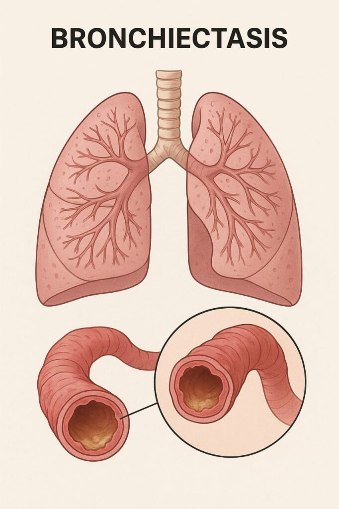
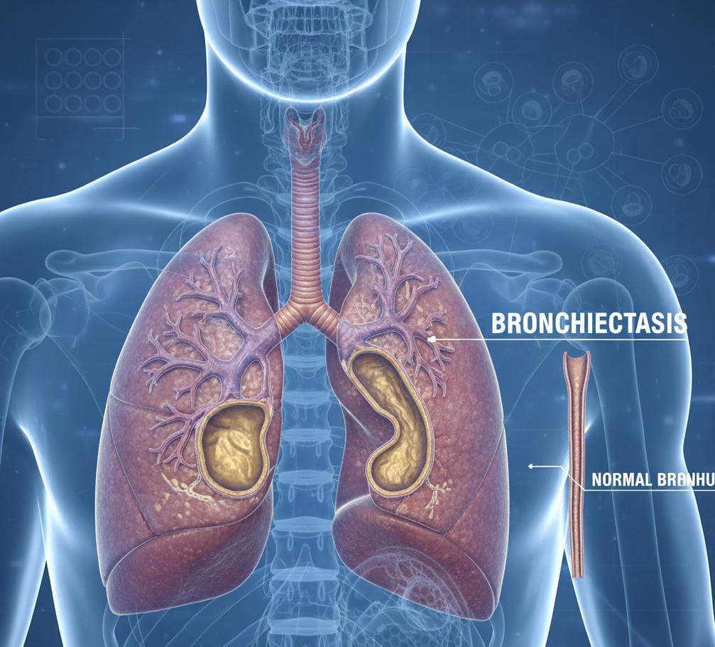
The word “bronchiectasis” comes from the Greek words “bronchus” (airway) and “ektasis” (expansion). In simple terms, it means abnormal and irreversible widening of the airways in the lungs.
In a healthy lung, the bronchial tubes carry air in and out of the lungs efficiently. However, in bronchiectasis, the walls of the bronchi become damaged, scarred, and thickened. This prevents the normal clearing of mucus, allowing bacteria to grow and cause repeated infections. Each infection can worsen the damage, creating a vicious cycle of inflammation and infection.
Bronchiectasis is a chronic lung condition where the airways (bronchi) become permanently widened and damaged due to repeated inflammation or infection. This structural damage causes mucus to build up, making it difficult for the lungs to clear bacteria and other harmful substances. Over time, this can lead to persistent cough, shortness of breath, and frequent respiratory infections.
Types of Bronchiectasis
There are generally two types of bronchiectasis, depending on the cause:
- Congenital Bronchiectasis
This form develops due to genetic or structural problems present at birth. It can occur as part of conditions such as:
Cystic Fibrosis (CF): The most common inherited cause of bronchiectasis.
Primary Ciliary Dyskinesia (PCD): A rare disorder affecting the cilia, the hair-like structures that help clear mucus from the lungs.
- Acquired Bronchiectasis
This type develops later in life due to infections, autoimmune diseases, or exposure to harmful substances. It is more common than congenital bronchiectasis and can affect people of all ages.
Causes
Bronchiectasis can result from a wide variety of factors that damage the airways. The main causes include:
- Repeated Lung Infections
- Frequent or severe infections such as pneumonia, tuberculosis (TB), whooping cough (pertussis), or measles can damage the bronchial walls and lead to bronchiectasis.
- Cystic Fibrosis
- A genetic condition that causes thick, sticky mucus to build up in the lungs, pancreas, and other organs. This mucus blocks the airways and leads to chronic infections.
- Immune System Disorders
- If the immune system cannot fight infections effectively, recurrent infections can cause chronic inflammation and scarring.
- Allergic Bronchopulmonary Aspergillosis (ABPA)
- An allergic reaction to the fungus Aspergillus can cause airway inflammation and damage.
- Primary Ciliary Dyskinesia
- This rare genetic disorder affects the movement of cilia in the airways, leading to poor mucus clearance.
- Obstruction of Airways
- A blockage caused by a foreign body, tumor, or enlarged lymph node can trap mucus and cause localized bronchiectasis.
- Autoimmune Diseases
- Conditions like rheumatoid arthritis, ulcerative colitis, and Crohn’s disease can also contribute to airway inflammation.
- Environmental Factors
- Long-term exposure to toxic gases, smoke, or pollution may damage the airways and contribute to bronchiectasis.
Pathophysiology (How Bronchiectasis Develops)
Bronchiectasis develops through a cycle of infection, inflammation, and airway destruction.
- Initial Injury: An infection or obstruction damages the bronchial walls.
- Inflammation: White blood cells release enzymes to fight infection, but they also harm the tissue.
- Mucus Accumulation: The damaged airways can no longer clear mucus effectively.
- Bacterial Growth: Trapped mucus becomes a breeding ground for bacteria.
- Further Damage: Repeated infections cause more inflammation, leading to irreversible dilation and scarring.
Symptoms
Symptoms of bronchiectasis can vary depending on the severity and extent of airway damage. Some people may have mild symptoms, while others experience severe, daily problems.
Common Symptoms Include:
- Chronic Cough: Persistent cough that lasts for months or years.
- Sputum Production: Thick, sticky, or foul-smelling mucus that may be yellow or green.
- Shortness of Breath: Especially during physical activity or exertion.
- Recurrent Chest Infections: Frequent need for antibiotics or hospitalization.
- Wheezing: Whistling sound when breathing.
- Chest Pain: Due to inflammation or infection in the lungs.
- Fatigue and Weakness: Ongoing infection and breathing difficulties can cause tiredness.
- Hemoptysis (Coughing up Blood): In severe cases, small blood vessels in the airways can rupture.
Complications
- Chronic respiratory failure
- Recurrent lung infections
- Collapsed lung (pneumothorax)
- Massive hemoptysis (severe bleeding)
- Cor pulmonale (right-sided heart failure due to lung disease)
Diagnosis
- Diagnosing bronchiectasis involves a combination of clinical evaluation, imaging tests, and lab investigations.
- Medical History and Physical Examination
- The doctor will ask about symptoms, past lung infections, family history, and exposure to irritants. Listening to the lungs with a stethoscope may reveal abnormal crackling sounds.
- Imaging Tests
- Chest X-ray: May show thickened or widened airways.
- High-Resolution CT (HRCT) Scan: The gold standard for diagnosis. It provides detailed images showing airway dilation and mucus plugging.
- Laboratory Tests
- Sputum Culture: Identifies bacteria or fungi causing infections.
- Blood Tests: To check immune function and detect underlying diseases.
- Sweat Chloride Test or Genetic Testing: To diagnose cystic fibrosis.
- Serum Immunoglobulin Levels: To rule out immune deficiencies.
- Lung Function Tests
- These measure how well the lungs are working and help monitor disease progression.
- Bronchoscopy
- A thin tube with a camera is inserted into the airways to look for blockages or collect samples for testing.
Treatment
- Airway Clearance Techniques
- Clearing mucus is a crucial part of managing bronchiectasis.
- Chest Physiotherapy (CPT): Involves clapping on the chest to loosen mucus.
- Postural Drainage: Using gravity to help drain mucus from different parts of the lungs.
- Positive Expiratory Pressure (PEP) Devices: Help open airways and move mucus.
- Oscillating Devices (e.g., Flutter Valve, Acapella): Create vibrations to loosen mucus.
- Medications
- Antibiotics: Used to treat or prevent bacterial infections.
- Bronchodilators: Help open the airways for easier breathing.
- Mucolytics: Thin the mucus so it can be coughed up more easily.
- Corticosteroids: Reduce airway inflammation (inhaled or oral forms).
- Vaccinations: Annual flu shots and pneumococcal vaccines reduce infection risk.
- Pulmonary Rehabilitation
- A structured exercise and education program helps improve lung capacity, endurance, and overall fitness.
- Surgery
- Surgical removal of the affected part of the lung may be considered if:
- The disease is limited to one area.
- Repeated infections occur despite medical therapy.
- Severe bleeding happens.
- Oxygen Therapy
- In advanced cases with low oxygen levels, supplemental oxygen may be prescribed.
Prevention
- Prompt treatment of respiratory infections
- Getting vaccinated against flu, pneumonia, and measles
- Avoiding smoking and secondhand smoke
- Managing chronic conditions like asthma and COPD effectively
- Regular medical checkups for early detection and intervention
Living with Bronchiectasis
Living with bronchiectasis can be challenging, but with proper care and lifestyle adjustments, it’s possible to maintain good health and prevent complications.
Tips for Daily Management:
- Follow your airway clearance routine daily.
- Take prescribed medicines as directed by your doctor.
- Track symptoms like cough frequency, sputum color, and breathlessness.
- Seek medical help early if infections worsen.
- Stay physically active to strengthen respiratory muscles.
- Stay away from pollution, smoke, and allergens.
Bronchiectasis in Children
Children can also develop bronchiectasis due to repeated lung infections or congenital conditions. Signs include:
- Persistent cough
- Failure to thrive
- Wheezing
- Frequent chest infections
Early diagnosis and treatment are essential to prevent long-term damage and support healthy lung development.