
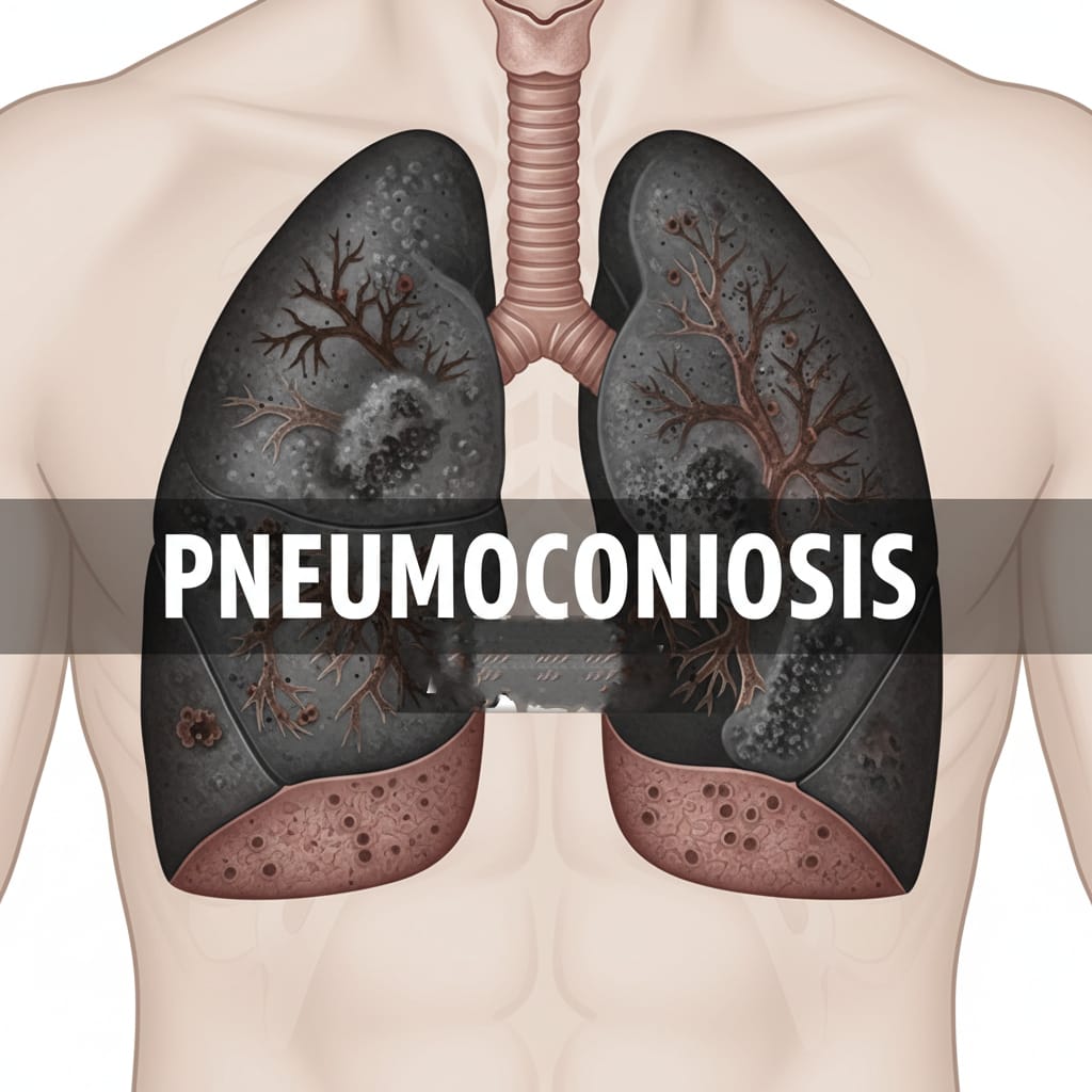
What is Pneumoconiosis?
Pneumoconiosis refers to a group of chronic lung diseases caused by inhalation of various types of dust, leading to lung tissue damage and fibrosis. The word originates from Greek:
- Pneumo = lungs
- Konis = dust
- -osis = disease
When dust particles enter the lungs and remain trapped, the body’s immune system reacts, causing inflammation and scar tissue formation. Over time, this scarring reduces lung elasticity, making it difficult to breathe and exchange oxygen effectively.
- Pneumoconiosis is not a single disease — it is a group of dust-related lung conditions, the most common of which include:
- Coal Workers’ Pneumoconiosis (CWP) — also known as Black Lung Disease
- Silicosis — caused by inhaling silica dust
- Asbestosis — caused by inhaling asbestos fibers
Pneumoconiosis: Causes, Symptoms, Diagnosis, Treatment, and Prevention
Pneumoconiosis is a serious occupational lung disease that develops after long-term inhalation of dust particles — especially mineral dust — in workplaces such as mines, factories, or construction sites. The condition leads to lung inflammation, fibrosis (scarring), and breathing difficulties. It’s considered one of the most common occupational lung diseases worldwide.
Types
- Coal Workers’ Pneumoconiosis (CWP)
- Common among coal miners, this type occurs due to long-term inhalation of coal dust.
- Simple CWP involves small black spots in the lungs.
- Complicated CWP or Progressive Massive Fibrosis (PMF) involves large scars, severely impairing breathing.
- Silicosis
- Caused by inhalation of crystalline silica dust, often from mining, quarrying, sandblasting, or stone cutting.
- Acute Silicosis develops rapidly (within months).
- Chronic Silicosis develops slowly over years.
- Accelerated Silicosis occurs within a few years of high exposure.
- Asbestosis
- Results from inhalation of asbestos fibers, used in insulation, shipbuilding, and construction.
- Long-term exposure can lead to lung fibrosis, pleural thickening, and increased risk of lung cancer and mesothelioma.
- Berylliosis
- Caused by exposure to beryllium dust or fumes, found in aerospace and electronics industries.
- It can cause chronic lung inflammation resembling sarcoidosis.
- Siderosis
- Occurs from inhaling iron oxide dust (common among welders).
- Generally mild, but prolonged exposure can cause fibrosis.
- Talcosis
- Develops due to inhalation of talc dust, often in talc mining or industrial processes using talcum powder.
Causes and Risk Factors
The primary cause of pneumoconiosis is inhalation of harmful dust particles that become trapped in the lungs. Over time, these particles trigger inflammation and scarring.
Common Causes:
- Long-term occupational exposure to mineral dust
- Poor ventilation at workplaces
- Lack of protective equipment (masks, respirators)
- Inadequate dust control systems
High-Risk Occupations:
- Miners (coal, silica, or asbestos)
- Construction workers
- Quarry workers
- Sandblasters
- Shipyard workers
- Welders and metal workers
- Pottery or ceramic industry workers
Other Risk Factors:
- Smoking (aggravates lung damage)
- Long duration of exposure
- Poor workplace safety regulations
- Weak immune system
Pathophysiology (How Pneumoconiosis Develops)
When dust particles enter the lungs:
- The macrophages (immune cells) try to engulf and remove them.
- Some dust particles are too resistant or sharp (like silica or asbestos).
- These particles damage macrophages, releasing inflammatory chemicals.
- Continuous inflammation leads to fibrosis (scarring) of lung tissue.
- The scarred tissue loses elasticity, making breathing difficult.
Over time, oxygen exchange becomes impaired, leading to shortness of breath, chronic cough, and respiratory failure in severe cases.
Signs and Symptoms
Symptoms may develop slowly over years, depending on exposure and dust type. In early stages, patients might not notice any problem.
Common Symptoms:
- Persistent dry cough
- Shortness of breath, especially during physical activity
- Chest tightness or pain
- Fatigue and weakness
- Bluish discoloration (cyanosis) due to low oxygen
- In advanced stages: respiratory failure, weight loss, finger clubbing
Specific Symptoms by Type:
- Silicosis: chronic cough, weight loss, fever, increased TB risk
- Asbestosis: chest pain, dry cough, crackling sound in lungs
- CWP: black sputum, progressive breathlessness
Complications of Pneumoconiosis
- Progressive Massive Fibrosis (PMF)
- Chronic Bronchitis and Emphysema
- Respiratory Failure
- Cor Pulmonale (right heart failure due to lung pressure)
- Increased risk of Lung Cancer (especially in asbestosis)
- Tuberculosis (in silicosis patients)
- Pleural effusion or thickening
Diagnosis
- Medical and Occupational History
- A detailed exposure history helps identify the source and duration of dust exposure.
- Physical Examination
- Doctors may listen for crackling sounds in the lungs or signs of breathing difficulty.
- Chest X-ray
A standard diagnostic tool showing small nodules, fibrosis, or masses typical of pneumoconiosis.
- High-Resolution CT (HRCT) Scan
Provides a more detailed image of the lungs, showing scarring patterns, nodules, and fibrosis.
- Pulmonary Function Tests (PFTs)
Measure lung capacity and airflow to assess the severity of damage.
- Blood Tests
May show reduced oxygen levels or rule out infections.
- Bronchoscopy and Biopsy
In some cases, a tissue sample from the lungs confirms the diagnosis.
Treatment
Currently, no cure exists for pneumoconiosis — once lung scarring occurs, it is irreversible. However, treatment focuses on slowing disease progression, relieving symptoms, and improving quality of life.
- Avoid Further Exposure
- The most critical step is to remove the patient from the source of dust exposure immediately.
- Medications
- Bronchodilators to ease breathing
- Corticosteroids (in certain cases) to reduce inflammation
- Antibiotics if infection develops
- Cough suppressants to reduce discomfort
- Oxygen Therapy
- Helps improve oxygen levels in the blood for those with chronic breathlessness.
- Pulmonary Rehabilitation
- Includes breathing exercises, physical training, and nutrition counseling to improve lung function and stamina.
- Vaccinations
- Patients should receive flu and pneumonia vaccines to prevent respiratory infections.
- Lung Transplant (in severe cases)
- For end-stage disease, a lung transplant may be considered if the patient is eligible.
Prevention
- Workplace Safety Regulations
- Enforce dust control systems (ventilation, wet drilling, dust suppression)
- Regular air quality monitoring
- Personal Protective Equipment (PPE)
- Workers should wear approved respirators or masks to prevent dust inhalation.
- Regular Health Check-ups
- Periodic chest X-rays and lung function tests help detect early signs of disease.
- Worker Education
- Training programs should inform workers about dust hazards and safe practices.
- Smoking Cessation
- Smoking increases the risk of lung damage — quitting helps preserve lung health.
- Proper Work Practices
- Rotating job duties, limiting exposure hours, and ensuring clean work areas reduce dust inhalation risk.